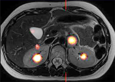
L-DOPA is used in the treatment of Parkinson’s disease; the compound crosses the blood-brain barrier and can be used to increase dopamine concentrations to temporarily relieve symptoms. Fluorodopa (F-DOPA) is a similar compound biochemically, with the main difference being the addition of a radioactive fluorine atom. TRIUMF’s nuclear-medicine group and its partners are working with L-DOPA and F-DOPA in aid of Parkinson’s research, and recently have started investigating its role in diagnosis of patients with neuroendocrine tumours. As well as being taken up by certain normal tissues within the body, F-DOPA also accumulates within neuroendocrine tumours (neoplasms which originate from the fetal neuroectoderm and contain secretory granules), including adrenal lesions such as phaeochromocytomas and paragangliomas. As a consequence of its radioactive decay, the F-18 atom attached to the DOPA molecule permits precise location of the tumours using a PET/CT scanner (see Figure 1). The synthetic organic chemistry performed to add the Fluorine-18 isotope can be done in only a limited number of places, including BC Cancer Agency and TRIUMF. As a result, these two organizations have been receiving referrals from all over Canada. TRIUMF has been doing work in the field of nuclear medicine for approximately 30 years now, and has become a driving force in it. Earlier this year, TRIUMF’s Mike Adam, along with doctors Daniel Levine, Daniel Metzger, Helen Nadel, Angelica Oviedo and Erik Skarsgard from BC Children’s Hospital, used F-DOPA and PET/CT imaging to diagnose a 16-year-old patient with a neuroendocrine tumor syndrome. Conventional magnetic resonance imaging showed three soft tissue tumours within the abdomen but failed to provide a unifying diagnosis. Even conventional nuclear medicine imaging with Iodine-123 labelled MIBG failed to clarify. However, F-DOPA PET/CT clearly depicted all three tumours and implied a common, unifying diagnosis, which was subsequently confirmed on genetic testing following successful surgical removal. This represented the first reported use of F-DOPA PET/CT for pediatric neuroendocrine syndrome imaging and was recently reported in the journal Pediatric Radiology. The combination of F-DOPA and PET/CT scanning has proved to be the most efficient method of detecting neuroendocrine tumours to date. In a test group of 14 patients, F-DOPA PET showed a sensitivity of 100%, compared to the traditional nuclear medicine imaging agent mIBG, labeled with Iodine-123, which had a sensitivity of 71%. Furthermore, F-DOPA PET/CT can be achieved in under two hours from injection to completion of imaging, whereas Iodine-123 mIBG imaging takes from 24 to 48 hours to complete. -- Written by Lindsay Davies, Communications Assistant |
Example of an F-DOPA PET/CT scan depicting the F-DOPA accumulation within the patient
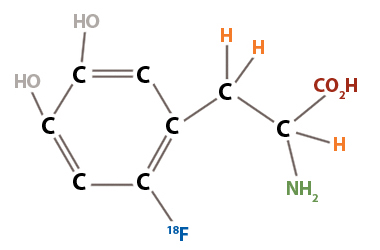
Figure 1: This illustration shows the structure of L-FDOPA, an aromatic amino acid. The neuroendocrine tumours metabolize the CO2H element (called decarboxylation), and as a result the F-18 is deposited in the same area, allowing the PET/CT scans to detect them and illustrate the location and size of the tumour |
Canada's particle accelerator centre
Centre canadien d'accélération des particules
Centre canadien d'accélération des particules